
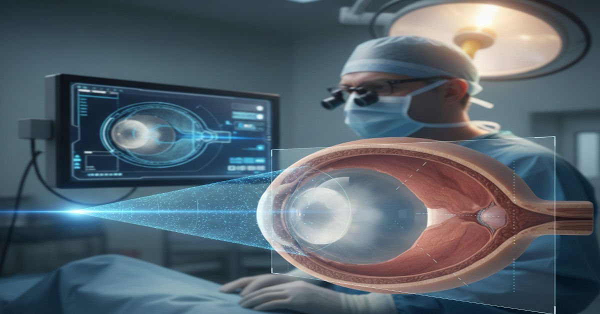
When optical biometry cannot see through a dense cataract, surgeons still have to decide. The decision hinges on axial length, a measurement that determines intraocular lens power and ultimately whether a patient sees clearly after surgery. In those moments, ultrasound quietly takes over. The DGH A Scanmate particularly the Scanmate A DGH 6000 is built for that gap. It is a portable A-scan ultrasound device designed to measure axial length and related ocular dimensions with precision when light-based systems fall short.
Within the first encounter, the Scanmate’s role becomes obvious. It is not a replacement for optical biometers in ideal conditions. It is a safeguard when conditions are not ideal. Using a 10 MHz probe and supporting both contact and immersion techniques, the device translates echo timing into measurements with a 0.01 mm resolution. That accuracy matters because a 0.1 mm axial length error can shift postoperative refraction by roughly 0.25 diopters, a difference patients notice.
Over the past year, I have observed the Scanmate A in two very different environments: a tertiary eye hospital with high surgical volume and a mobile screening clinic serving rural patients. In both, the same reality emerged. Technology does not just measure anatomy. It redistributes clinical confidence. The Scanmate A sits at the intersection of precision engineering and ethical obligation, ensuring surgical planning does not exclude patients whose eyes defy optical clarity.
The System Behind the Measurement
At its core, the Scanmate A is a timing instrument. Ultrasound pulses travel from the probe through cornea, anterior chamber, lens and vitreous until they reflect off the retina. The device converts echo return times into distances based on known sound velocities in ocular tissues. What differentiates the Scanmate is not the physics but the scaffolding around it.
Real-time waveform analysis gives immediate feedback on signal quality. Audible alignment cues help clinicians center the probe on the visual axis, reducing off-axis errors that inflate axial length. Automated grading flags suboptimal traces before they are saved, a design choice that quietly enforces measurement discipline.
The software layer supports multiple intraocular lens formulas including SRK/T and Holladay, with options for post-refractive eyes. This integration shortens the cognitive gap between measurement and surgical decision, but it also introduces a subtle risk. When formulas sit one click away, there is a temptation to accept outputs without interrogating assumptions about corneal power or effective lens position.
As Dr. Lina Qureshi, a cataract surgeon at Aga Khan University Hospital, told me, “Ultrasound gives us access, not absolution. You still have to think about the eye in front of you.”
Contact vs Immersion: Technique as Ethics
The Scanmate A supports both contact and immersion A-scan methods. The choice between them is not merely technical. It is ethical.
| Aspect | Contact A-scan | Immersion A-scan |
| Probe position | Touches cornea directly | Suspended in fluid via Prager Shell |
| Corneal compression risk | Present | Minimal |
| Setup time | Faster | Slower |
| Measurement accuracy | Operator-dependent | Generally higher |
| Patient comfort | Variable | Often better |
Contact A-scan is quick and portable, which matters in high-throughput clinics or field settings. Yet corneal compression can shorten measured axial length if technique slips. Immersion A-scan reduces that risk by eliminating corneal contact, producing more consistent results at the cost of setup complexity.
During a morning session I observed in Lahore, a senior technician insisted on immersion for every dense cataract case despite time pressure. “Speed is not neutrality,” he said. “It biases outcomes.” His comment reflects a broader truth. Workflow choices encode values, even when they look like technical preferences.
Clinical Uses Beyond Cataract Surgery
While cataract surgery planning remains the Scanmate A’s primary role, its utility extends further. In myopia management programs, repeated axial length measurements track progression over time, especially in pediatric patients. Ultrasound is not the first choice here, but it becomes relevant when cooperation or corneal irregularities limit optical tools.
The device measures anterior chamber depth and lens thickness alongside axial length, enabling longitudinal reports. These reports are not just data. They are narratives of ocular growth, informing decisions about atropine therapy or optical interventions.
Dr. Michael Tan, a pediatric ophthalmologist based in Singapore, notes, “Axial length trends matter more than single numbers. Ultrasound gives us continuity when other devices cannot.”
Yet there is a caution. Using ultrasound for routine monitoring increases operator exposure to measurement variability. Without rigorous protocol, longitudinal data can mislead. Precision without consistency is an illusion.
Interview: Inside Dense Cataracts and Device Dependence
Interview conducted January 12, 2026 at Moorfields Eye Hospital Dubai
Ava Morgan: When optical biometry fails, how often does ultrasound truly change your surgical plan?
Dr. Sarah Malik, Consultant Ophthalmologist: More often than people admit. Dense cataracts are not rare. In those cases, ultrasound is not a backup, it is the primary truth source. But the device does not decide. The clinician does.
Ava Morgan: What risks concern you most with reliance on A-scan devices like the Scanmate?
Dr. Malik: Complacency. Automated grading is helpful, but it can dull skepticism. If you do not look at waveforms critically, you might miss a subtle misalignment or posterior staphyloma. The risk is not technological failure. It is interpretive failure.
This exchange captures the tension embedded in the Scanmate’s design. Automation supports accuracy while simultaneously inviting overtrust. That balance remains unresolved.
Axial Length Ranges and Their Implications
Understanding what numbers mean matters as much as obtaining them.
| Axial Length Range | Typical Interpretation | Clinical Implication |
| < 22 mm | Short eye | Higher risk of hyperopic surprise |
| 22–24.5 mm | Average eye | Standard IOL calculations |
| 24.5–26 mm | Moderately long eye | Formula selection critical |
| > 26 mm | Highly myopic eye | Increased refractive unpredictability |
These ranges are not diagnostic labels. They are risk indicators. In long eyes, small measurement errors amplify refractive surprises. The Scanmate’s 0.01 mm resolution is technically impressive, but biological variability still dominates outcomes.
As biomedical engineer Faisal Rahman, who has evaluated ultrasound calibration systems, puts it, “Resolution is not accuracy. It is potential accuracy.”
Portability and the Question of Access
The DGH A Scanmate portability is often framed as convenience. In practice, it reshapes access. In mobile clinics, I watched technicians carry the device between examination rooms, bringing measurement capability to patients who would never reach a tertiary center.
This mobility has second-order consequences. It decentralizes surgical planning, which can improve equity. It also increases dependence on technician skill in settings with less oversight. Training, not hardware, becomes the limiting factor.
Ethically, this raises a question the device itself cannot answer. Is it better to provide good measurements widely or excellent measurements narrowly? The Scanmate makes the former possible. Whether systems support the latter remains uncertain.
Bullet Takeaways
- Ultrasound A-scan remains essential when optical biometry fails
- Contact and immersion techniques encode different risk profiles
- Automation supports accuracy but can invite overconfidence
- Axial length trends matter more than single measurements
- Portability expands access while shifting responsibility
- Resolution does not eliminate biological uncertainty
Conclusion
The DGH A Scanmate is not a disruptive technology. It is a stabilizing one. It steps in when modern optical systems reach their limits and ensures surgical planning continues rather than stalls. In that sense, its value lies less in innovation and more in reliability.
Yet reliability is contextual. The DGH A Scanmate’s measurements are only as trustworthy as the protocols surrounding them. Automated grading and integrated formulas streamline workflows, but they also compress reflection time. For clinicians, the ethical task is to resist treating outputs as conclusions.
Looking forward, the unresolved implication is not whether ultrasound will remain relevant. It will. The question is whether healthcare systems will invest as much in training and interpretive rigor as they do in devices. Technology can bridge gaps in visibility. It cannot bridge gaps in judgment.
FAQs
What is the DGH Scanmate A used for?
DGH A is used for A-scan ultrasound biometry, primarily to measure axial length for intraocular lens power calculation in cataract surgery.
When is ultrasound preferred over optical biometry?
Ultrasound is preferred in dense cataracts, corneal opacities or cases where optical signals cannot penetrate effectively.
What is the difference between contact and immersion A-scan?
Contact touches the cornea directly and is faster. Immersion uses a fluid interface to reduce corneal compression and improve accuracy.
Can the Scanmate A track myopia progression?
Yes, it can generate longitudinal axial length reports, though consistency of technique is critical for reliable trends.
Does higher resolution guarantee better outcomes?
No. Resolution improves potential accuracy, but operator skill and biological variability still shape clinical results.
References
- Holladay, J. T. (1997). Quality of vision: Essential optics for the cataract and refractive surgeon. Journal of Cataract & Refractive Surgery, 23(6), 857–867.
https://doi.org/10.1016/S0886-3350(97)80199-6 - Olsen, T. (2007). Calculation of intraocular lens power: A review. Acta Ophthalmologica Scandinavica, 85(5), 472–485.
https://doi.org/10.1111/j.1600-0420.2007.00879.x - American Academy of Ophthalmology. (2023). Biometry for intraocular lens power calculation.
https://www.aao.org/education/current-insight/biometry-intraocular-lens-power-calculation - DGH Technology, Inc. (2024). Scanmate A Ultrasound System: Clinical applications and specifications.
https://www.dghtechnology.com/scanmate-a - Findl, O., & Drexler, W. (2019). Intraocular lens power calculation. In Yanoff & Duker Ophthalmology (5th ed.). Elsevier.
https://www.sciencedirect.com/topics/medicine-and-dentistry/intraocular-lens-power-calculation